When you get a bleed or feel pain in a joint or another part of your body, your comprehensive care team (CCT) needs to get a closer look at what is happening inside your body. The sooner your team finds out the extent of your bleed, the sooner they will know the best way to treat your bleed.
In hemophilia, radiological imaging is often used to get a more detailed picture of the internal structures inside your body, such as your joints. This is also helpful when you bleed into a tight space like your organs, spine or head.
Imaging also provides useful information on how the bleed is affecting other areas, such as the synovium, cartilage and bones. These areas may be affected from a bleed, even if you do not feel any pain there.
To get more information on your bleed, one or multiple types of imaging techniques may be used. These include X-rays, ultrasound, computed tomography (CT) and magnetic resonance imaging (MRI).
X-rays
An X-ray is a type of radiation that passes through the body. It’s a quick way for your doctor to visualize the internal structures of your body. This includes the size, shape, and location of the bones and certain organs.
How is the X-ray taken?
You will wear a hospital gown and be asked to stand next to the X-ray film in the hospital radiology department. The technician will ask you to hold still for two or three seconds so the picture doesn’t blur. The X-ray machine is turned on for a fraction of a second. Beams of X-rays pass through the part of your body to make a picture on the film. Usually an X-ray is taken from the back and then the side. Like cameras, most X-ray imaging is now digital, so images are available instantly. The image will show patches of white and black. The densest parts of your body, such as bones, will show up as white patches, while less dense parts are usually grey. Any plates or screws that doctors may have placed in your body will also show up in the X-ray. Since they are denser than your bones, plates or screws will appear whiter in the X-ray.
Does an X-ray hurt?
No, you won’t feel a thing. There is very little radiation released through the X-ray so it won’t cause damage to the body.
Ultrasound
When you have a bleed, doctors need to find out whether there is bleeding into the joint or surrounding structures. An ultrasound uses sound waves to take pictures of the inside of your body. These pictures give your doctor information about the size, shape, and texture of the body part being scanned.
A sonographer (ultrasound technologist) performs your test. A sonographer is an expert in the use of ultrasound machines. The sonographer will give a report of the scan to your doctor.
How long an ultrasound scan takes depends on the location and extent of the injury. It typically takes between 20 and 45 minutes.
During an ultrasound
The ultrasound is taken inside a scanning room. The scanning room has a bed, an ultrasound machine and a screen that shows the pictures from the ultrasound.
The sonographer uses a small hand-held probe to take pictures of the body. The sonographer will put a warm gel on the probe or your skin. The gel feels like warm soft cream and does not hurt. After you lie down on the bed, the probe is gently placed on your skin over the area to be scanned.
As the probe moves over your body, a moving picture will appear on the ultrasound screen. The sonographer takes many pictures during the test.
Magnetic resonance imaging (MRI)
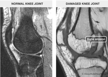
Magnetic resonance imaging (MRI) shows the internal structures inside your body in tremendous detail, including tiny structures like your veins, arteries and nerves.
An MRI machine does not use radiation or X-rays. A magnet, radio signals and a computer are used to create the pictures. It scans the area of your body in “slices” to produce images. They are also called cross-sectional images. For example, if you’re getting an MRI on your brain, each “slice” shows a different level of the brain. These images or scans are interpreted by a radiologist.

The person who performs your MRI is called an MRI technologist. No one is allowed to go near an MRI machine unless they have first been screened for magnetic metals inside or on their bodies, such as piercings. This is important because they could get stuck to the machine, which could damage the machine, or create an image that is too blurry to read, it could also be very dangerous.
What happens during an MRI test?
To produce the image, your entire body goes into the MRI machine. Coils inside the machine focus and zoom in on specific parts of your body. If you get an MRI, your doctors are looking for any early signs of joint damage. This way, you can learn how to prevent the damage on those joints from becoming worse.
How long does it take?
An MRI scan usually takes 30 to 60 minutes, but it may take longer. An MRI can be done on any area of the body.
Does it hurt?
An MRI scan is fairly noisy but does not hurt.


